The paper was printed also in: ...S. M. Kaczmarek, R. Jabłoński, I. Pracka, G. Boulon, T. Łukasiewicz, Z. Moroz and S. Warchoł, „Radiation Defects in SrLaGa3O7 Crystals Doped With Rare-Earth Elements”, Nuclear Instruments and Methods in Physics Research Section B, Beam Interactions with Materials and Atoms, B142, 1998, 515-522
|
|
|
Biuletyn WAT
Influence
of ionizing radiation on SrLaGa3O7 single crystals doped
with rare-earth elements
SŁAWOMIR MAKSYMILIAN KACZMAREK RYSZARD JABŁOŃSKI*
IZABELLA PRACKA* GEORGES
BOULON**
TADEUSZ ŁUKASIEWICZ*** ZBIGNIEW MOROZ****
STANISŁAW WARCHOŁ***** KRZYSZTOF STĘPKA
*
Institute of Optoelectronics, MUT, 00-908 Warsaw, ul. S. Kaliskiego
2, Poland
* Institute of Electronic Materials Technology., 01-919 Warsaw, ul. Wólczyńska
133, Poland
**
Laboratoire de Phys.-Chimie des Materiaux Luminescentes, 69122 Villeurbanne
Cedex, France
*** Institute of Physics MUT, 00-908 Warsaw, ul. S. Kaliskiego 2, Poland
****
Soltan Institute of Nuclear Sudies, 05-400 Świerk, Poland
*****
Institute of Chemistry and Nuclear Technics, 02-415 Warsaw, ul. Dorodna
16, Poland
Abstract.
Influence of g-rays
from a 60Co (1,25 MeV) source and 26 MeV protons on the absorption
and luminescence of SrLaGa3O7 single crystals doped with
Nd, Dy and Pr was studied. Color centers, which appeared after an irradiation (absorbed
dose: 105 Gy), shift the absorption edge towards the longer
wavelengths by a few hundreds nm (for a rod length about 36 mm). Measurements of
the ESR spectra before and after gamma irradiation were also performed. They
show spectra with a spin of S = 1/2, g|| = 1.9838(5) and g^
= 2.0453(5), that can be attributed to the Ga2+ centers formed
according to the pattern Ga3++e-®Ga2+.
Optical output measurements for Nd doped SLGO laser rods showed
some improvement of laser emission at 1.06 mm for strongly defected rod. This is due to the same paramagnetic Ga2+
color centers that arise in SLGO crystals after g-
or proton irradiation.
Keywords:
materials engineering, color centers, ionizing radiation, luminescence
Universal
Decimal Classification:
620.1
The
melilites (ABC3O7: A = Ca, Sr, Ba; B = La, Gd; and C = Ga,
Al) are layered compounds which consist of vertex-sharing tetrahedral units
linked together forming distorted planar networks with a five-membered ring
pattern. Alkaline or rare-earth cations are sandwiched between these layers and
located in sites with a very distorted square Archimedean antiprism
configuration [1, 2]. Single crystals of SrLaGa3O7 (SLGO)
exhibit the highest structural homogeneity among these compounds. They appear to
be promising active materials for the design of all-solid-state lasers [3, 4].
They exhibit, however, strong changes in absorption and luminescence spectra
after irradiation by ionizing particles. Irradiation causes a very large
additional absorption near the short-wavelength absorption edge, which depends
on radiation dose and on sample thickness. These changes influence properties of
lasers in which the SLGO crystals are used as the active material.
In the
present work we attempted to study more detailed influence of gamma and proton
radiation on optical properties of Nd, Dy and
Pr ion doped SLGO crystals.
The
samples of SLGO crystals doped with Nd (5 at. % and 10 at. %), Pr (1 at. %, 0,5
at. %) and Dy (1 at. %, 0,5 at. %) were grown by the Czochralski method in
iridium crucibles. The detailed description of the applied growth process is
presented elsewhere [5-6].
Gamma
irradiation from a 60Co source at a dose rate of 1.5 Gy/sec up to an
absorbed dose of 106 Gy was applied. For proton irradiation the beam
from the cyclotron C-30 was used. The average energy of protons was about 26 MeV
and fluencies varied between 1013 and 1016 particles/cm2.
After
each proton irradiation gamma lines were measured and types of nuclear reactions
defined. For protons with a dose of 1016 protons/cm2, they
are presented in Table 1.
Table 1.
Energies
of gamma lines and types of nuclear reactions for Dy3+ doped SLGO
crystal (0,5at.%) irradiated by protons with a dose of 1016 protons/cm2.
|
Eg
(keV) |
Nuclear
reaction |
T1/2
(days) |
|
1 |
2 |
3 |
|
165 |
|
137,7 |
|
Ex=33,
33.4, 37.8, 38.7 |
|
|
|
|
|
3 |
|
387 |
|
|
|
484 |
|
|
|
|
|
106,6 |
|
814
(1835-1022) |
|
|
|
898 |
|
|
|
1325
(1836-511) |
|
|
|
1836 |
|
|
|
2734
(sum peak) |
|
|
|
|
|
244 |
|
1116 |
|
|
|
|
|
270 |
|
1078 |
|
|
Samples
of SLGO crystals doped with Nd3+, Pr3+ and Dy3+
diameter of 10 mm and 1-2 mm thick were cut out perpendicularly to the growth
axis in the plane (111) from the most homogeneous part of crystals. After
optical polishing of both ends, the crystals were examined with a Mach-Zehnder
interferometer. Samples in form of a rod of 4 mm diameter and length of about 36
mm were also investigated. To obtain absorption coefficients in the range of
200-1100 nm, transmission spectra
of the samples were measured before and after g
or proton irradiation using a LAMBDA-2 Perkin-Elmer spectrometer.
Values
of additional absorption (DK factors) caused by the irradiation were calculated from the formula:
![]() (1)
(1)
where
l
stands for wavelength, d for the sample thickness, T1 and T2
for transmission of the sample before and after gamma irradiation, respectively.
Fluorescence
and excitation spectra were obtained using an ILA - 120 3 W argon ion laser. The
spectra were recorded using a GDM-1000 monochromator with dispersion of 11 cm-1/mm
and detected by a RCA C-31034-02 cooled AsGa photomultiplier. For data
aquisition the SR 400 photon counting system, controlled with a PC computer, was
used.
For the
thermoluminescence studies unpolished samples were prepared, with a thickness
lower than 1 mm and diameter up to 6 mm. Thermoluminescence was measured for
crystals 'as grown' and gamma irradiated in
the temperature range from 70 to 400 oC, by means of a "
carousele " analyzer WAWA-TLD RA'95 installed in the Institute of Chemistry
and Nuclear Technique in Warsaw.
The
samples, typically of 3,5´3,5´2
mm, were measured in a BRUKER ESP-300 ESR spectrometer (X-band). The
spectrometer was equipped with helium flow cryostat type ESR-900 Oxford
Instruments. The ESR lines were observed before and after gamma exposure of 105
Gy dose in the temperature range from 4 to 300 K and microwave power from 0,002
to 200 mW. Moreover, the above investigations were performed for crystals
annealed in air at 800 oC for three hours.
Four
rods cutted out from the same crystal were investigated. Measurements were made
in naturally air-cooled head for the laser system pumped by xenon flash-lamp
with energies from 5 to 50 J and a filter inside of the head cutting wavelength
up to 350 nm. Laser rods were put into the plane-parallel laser cavity 24 cm
long, made of messing covered with gold. Transmissions of output mirrors were
equal to 8%, 19.5% and 36,6%, respectively, for 1,06 mm.
The rods had no AR coatings on their end faces. The laser light was detected
with high-sensitive HgCdTe photoconductor and time characteristics of the lamp
were observed by Si photodiode. The energy of laser pulses was measured by
Gen-Tec radiometer with ED-500 gauge head.
All the
rods were investigated subsequently: as grown, irradiated by gamma’s of 103-106
Gy, annealed in air at 1200 oC for three hours, polished (end faces
only) and again gamma-irradiated with 103 - 106 Gy.
The
thermoluminescence measurements of
the Nd: SLGO (5 at. %) crystals
show, that in the crystal 'as grown' ( curve 1, Fig 1) only one defect exist
with maximum at 319 oC. In the same, but gamma irradiated crystal,
measured just after g-irradiation
with a dose of 103 Gy (see Fig. 1. curve 2), two defects are seen: at
110 oC and 260 oC, respectively. The defect at 110 oC
is the new one introduced to the crystal by gamma irradiation.
As
seen from Fig. 1 changes for Nd: SLGO crystals in a thermoluminescence spectrum
caused by g-irradiation
rapidly relaxes and stays almost the same for both doses: 103 Gy (measured
just after g-exposure)
and 105 Gy (measured after 1,5 month from g-exposure, see Fig. 1. curves 2 and 3). Moreover, the maximum of the
thermoluminescence shifts with time.
Additional absorption (AA) bands observed in the Nd: SLGO (5 at. % and 10 at. %) and Pr: SLGO (1 at. %, 0.5 at. %) crystals after gamma irradiation with a dose of 105 Gy and in the Dy: SLGO (1 at. % and 0.5 at. %) crystals with a dose of 106 Gy are shown in Fig 2. As seen, in SLGO crystals doped with Dy, Nd and Pr gamma induced AA bands appear at about 290 nm and 380 nm, but the first one is much stronger than the second one. The first AA band intensity and location depend on the gamma dose and on the kind of the dopant. With the grow of the Pr or Dy concentration, the intensity of one of these bands decreases, but with the increase of the Nd concentration, it also increases. Therefore, intensity of this band as a function of dopant concentration strongly depends on the type of dopant.
Fig. 1. Thermoluminescence
curves of Nd: SLGO (5at.%) ‘as grown’ crystal before ( AG -1) and after g:
103Gy
(2 -just after
irradiation)
and 105 Gy
(3 - 1,5 month later)
irradiation.
Moreover,
after gamma irradiation of a thin SLGO sample the shift of about 50 nm of a
short-wave absorption edge was observed. The value of the shift strongly depend
on gamma dose.
In Fig 2
one can see also the AA bands for the Nd: SLGO (5 at. % of Nd3+) rod
(curve 3), compared with those for a 2.1 mm plate (curve 2). Shift of the
short-wave absorption edge is caused by a large value of rod length (36.16 mm)
and a scattering nature of absorbing centers. Fig. 3 presents changes in
transmission and absorption spectra of “as grown” Dy: SLGO crystal (1 at. %)
(curve 1) after subsequent: gamma irradiation of 106 Gy (curve 2),
thermal annealing in air at 1200 oC for
three hours of the
irradiated sample (curve
3) and gamma exposure of 103 Gy of the annealed sample (curve 4). One
can see, the shift of short-wave absorption edge and two different radiation
defects can be observed. First one, with a maximum at 380 nm, is connected
probably with recharging effect of oxide vacancies while second one, with
maximum at 290 nm, is a new radiation defect.
Fig.
2 Additional absorption bands of Dy, Pr and Nd doped SLGO crystals after gamma
irradiation with doses of 105 (Pr and Nd:SLGO) and 106 Gy
(Dy:SLGO).
The first of these
defects (380 nm) appears mainly after previous annealing of the crystal and
following g-exposure.
It is related to curve 5 from Fig. 2 (Pr: SLGO (0,5 at. %)) as well as to curve
4 from Fig. 3 (Dy: SLGO (1 at. %)).
Fig. 3. Changes in
transmission and absorption spectra of ‘as grown’ Dy: SLGO (1at.%) crystal
(1) after subseqent: gamma irradiation with a dose of 106 Gy (2),
annealing in air at 1200oC for 3 h (3) and gamma exposure with a dose
of 103 Gy (4).
The first of these
defects (380 nm) appears mainly after previous annealing of the crystal and
following g-exposure.
It is related to curve 5 from Fig. 2 (Pr: SLGO (0,5 at. %)) as well as to curve
4 from Fig. 3 (Dy: SLGO (1 at. %)).
The
Dy: SLGO crystal (0.5 at. %, d = 2.91 mm, d - sample thickness) was also
irradiated by protons (1013 - 1016 particles/cm2).
The AA bands were obtained at about 270 nm and 370 nm (the first maximum on the
level of 23 1/cm). The shift of the absorption
edge after proton exposure
was about 50 nm. This is essentially the
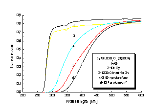
Fig.
4. Transmission spectra of gamma and proton exposure Dy: SLGO (0,5at.%) crystal
for doses up to 1016 protons/cm2
Shifts of
the short-wave absorption edge for Dy: SLGO
(0.5 at. %) crystal after gamma and proton exposures are seen together in
Fig. 4. The as grown Dy: SLGO crystal (curve 1) was firstly gamma irradiated
with a dose of 105 Gy (curve 2), then annealed at 1200 oC
in air for three hours (curve 3) and next subsequently irradiated with protons
with doses: 3*1013, and 1,12*1016
protons/cm2 (curves 4, and 5). One can see that the shifting due to
gamma irradiation is greater than that one due to protons 3*1013 cm-2.
Much
more clearly this situation is described in
Fig. 5, where for different gamma doses, changes of the transmission measured
for the Nd: SLGO (5 at. %) rod of the length
of 36.16 mm are shown. This rod (curve 1) was subsequently irradiated by
gamma’s with 104 Gy (curve 2), annealed at 400 oC for 3
hours, irradiated with 105 Gy (curve 3), annealed at 1200oC
for three hours (curve 4) and, finally, irradiated with 105 Gy (curve
5). As the absorption edge, the wavelength was taken for which the transmission
value reach the level of 0.001.
As seen
from the figure, the short-wave absorption edge became shifted with the increase
of the radiation dose towards the longer wavelengths. This shift, for the rod of
about 36 mm length and 106 Gy, may even be as large as 120 nm. As
seen from Fig. 5, after annealing the rod at 1200 oC, absorption edge
return to position before irradiation (curves 1 and 4). Moreover, for the same
values of gamma doses but different conditions of irradiation (irradiation after
annealing at 400 oC and irradiation after annealing at 1200 oC
with a dose of 105 Gy - curves 3 and 5) absorption edge is also the
same.
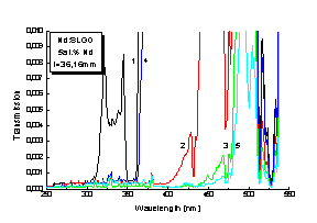
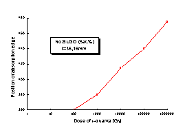
Fig.
6. Change of absorption edge position in Nd: SLGO (5at.%) crystal after g-irradiation
with doses from 102 to 106 Gy
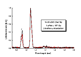
Fig.
7. Luminescence spectrum for Nd: SLGO (5at.%)crystal after (1) and before (2) g-irradiation
with dose of 103 Gy
Gamma
irradiation with different doses of 4 different rods but of the same length and
thickness, cut from the same crystal was performed. The change of the short-wave
absorption edge as a function of gamma dose is shown in Fig. 6. It is seen from
the Fig. 6., that this change is linear as a function of the dose. Similar
dependence was observed for plates cut from
2 mm Nd: SLGO and Dy:
SLGO crystals, but
the shifts were smaller (a maximum about 50 nm). This suggest that the color
centers responsible for this shifting are of a scattering type. Similar
investigations performed for Pr: SLGO rod with f = 4 mm and l = 39.12 mm gave the shift of about 200 nm, for 106
Gy gamma’s.
Fig. 7 shows relative changes in a luminescence of the Nd: SLGO (5 at. %)
crystal before and after the 105 Gy gamma exposure. Small decrease in relative values of a luminescence close to
l
= 910 nm can be
noticed.
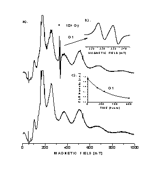
Fig.8
. ESR spectrum for SLGO crystal before and after g-irradiation
with the dose of 105 Gy. 8a). ESR spectra before and after g-exposure,
8b). G1 defect in SLGO crystal and 8c). Time quenching of G1 defect. Fitting was
performed for the curve: y=y0+A1*exp(-(x-x0)/t),
where:x0=0, y0=0.06245, t=264h, A1=0.86 and csqr=7.516*10-5
Its angular dependence for (001) plane is depicted in Fig. 9. The
g-factor varies in the range of 2,0045 - 2,044. The same type of angular
dependences for (100) plane was also observed.
These
lines are observed at temperatures from 4 to 300 K but above 227 K due to line
broadening, only isotropic single line is observed. The above mentioned lines
appear in SLGO crystal after gamma irradiation independently on the kind of
impurity, also in the undoped SLGO crystal.
After
annealing the crystal in air at 800 oC for three hours these lines
disappear. Moreover, after 1 month of storage the sample at room temperature the
intensity of ESR lines generated after irradiation decreases about 10 times
(Fig.8. - curve: ESR intensity as a function of a time) which gives the
life-time of the observed center of 264 hours.
For
three of four Nd: SLGO rods, (‘good rods’ with a low value of laser emission
threshold at 1.06 mm), a decrease
of optical output after g-exposure and further after thermal annealing was observed as it is shown
in Fig. 10. Values of slope efficiencies of Nd: SLGO lasers are described also
as a number near each optical output curve. The results of the investigations of
all the rods before and after g-irradiation
of ‘as grown’ crystals are presented in Ref. [7].
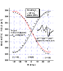
Fig. 9. Angular dependences of g-irradiation defect G1 in (001) plane of SLGO crystal. Small picture inside the figure show ESR spectrum for [110] direction. P denotes phosphorus lines (signal lines)
The results
for the fourth rod (‘bad rod’ with a high value of laser emission threshold
at 1.06 mm)
are presented in Fig. 11., which shows an increase of output energy after gammas
exposure and again a decrease after thermal annealing.
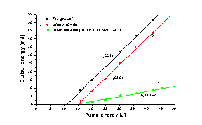
Fig. 10. Optical output of a ‘good’ Nd: SLGO (5 at. %) laser emitting at 1.06 mm, for different kinds of processing: 1-’as grown’, 2-g-105 Gy of ‘as grown’ rod and 3-annealing the rod in air at 1200 oC for 3h. Numbers near curves denote slope efficiencies of the laser
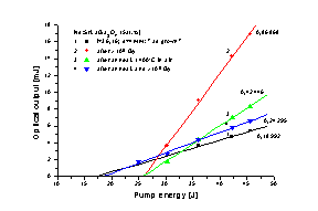
Fig.
11. Optical output of a ‘bad’ Nd: SLGO (5 at .%) laser emitting at 1.06 mm,
for different kinds of processing: 1 - ’as grown’, 2 - g-105
Gy of ‘as grown’ rod, 3 - annealing the rod in air at 1200 oC for
3h and 4 - g-105
Gy - irradiation of previously annealed the rod
It was
stated in Ref. [7] that in Nd: SLGO (5 at. %) lasers after gamma irradiation one
can obtain an improvement of their optical
output, provided that these crystals had been strongly defected.
Probably,
the output energy of a laser increases as a result of appearance of color center
(CC), shifting the absorption edge and, in this way, lowering the absorption in
the UV region (lowering the optical losses) and also changing the amplitude
relations of the luminescence.
In Fig.
1. one can see this CC having its maximum at 110 oC and relaxing (curves
2 and 3) with time. Second CC with a maximum at 260 oC that arises
also for unirradiated crystal is connected probably with an oxide vacancy in
SLGO structure and arises also in undoped crystal. The same behavior show also
Nd:BaLaGa3O7 [8], Cr:SrGdGa3O7 and
also SLGO crystals doped with Dy and Pr ions. In Figs 2. and 3. two the same
CC’s are seen in absorption spectra. The first one, with a maximum at about
290 nm, is connected with a paramagnetic defect generated in SLGO crystal by g-irradiation
and causes a strong absorption (tens of 1/cm) in the region of the absorption
edge. The second one, with a maximum at about 380 nm, is connected probably with
a recharging effect of oxide vacancy. It is observed especially after g-
or proton irradiation of the crystal previously thermally annealed.
The
intensity and location of the first center strongly depend on the kind and
concentration of a dopant. For the neodymium increase of
the intensity is observed with an increase of Nd concentration in SLGO
crystal, while for Pr and Dy a decrease of intensity is observed.
The
intensity depends also on radiation dose. With the growth of dose the value of
the intensity also increases.
A great
value of the AA arising after g- or proton-irradiation near short-wave absorption edge causes shifting
of the edge, which for a gamma dose of 106 Gy can have value of 50
nm. This shifting depends on gammas or protons dose and on thickness of
investigated sample. This suggests that the CC responsible for this shifting is
of a scattering type.
The
dependence of the shifting on gamma absorbed dose for Nd: SLGO crystal is
presented in Fig. 6. As one can see, it have linear character as a dose function
and for rods with a length of 36 mm can have a value of 200 nm.
Arising
of this CC in Nd: SLGO crystal causes small changes in luminescence spectrum
seen at a wavelength of 910 nm in Fig. 7.
Improvement
of the laser emission in a ‘bad’ Nd: SLGO rod at 1,06 mm
which is seen in Fig. 11 is probably due to characteristic radiation defect,
that is connected with a greater than in Nd: YAG increase of AA value in the
range of absorption pump spectrum. For Nd: YAG rod we have obtained Dkmax = 0.7 [1/cm], while for Nd: SLGO Dkmax
= 2 [1/cm] both in UV range of absorption spectrum for a gamma dose of 105
Gy.
Growth
of the laser emission threshold after thermal annealing, which is seen also in
figures 10 and 11, is due to the fact that for the temperature of 1200 oC
some dopants contained in the crystal are oxidized. In this way transparency for
both end faces of a rod, as well as its side surface, changes essentially. End
faces were polished after annealing process in contrary to side surface. The
decrease of the laser slope efficiency after thermal annealing process of the
laser rod can be explained by arising in the crystal CC with a maximum at 380 nm
after g- or proton
irradiation.
Fig.12a.
illustrates crystallographic positions of La+
, Ga3+, Sr3+ and O2- ions in the SLGO lattice (c-axis
projection). In this figure one can see six T1, T2...T6 tetrahedra of GaO42-
type.
Fig.
12b. shows complexes of (Ga-O)1- arising in SLGO structure (T1
tetrahedron) after g-
or proton irradiation.
The
obtained results can be explained by means of the following process: Ga3+
ion captures the electron which was knocked out from O2- ion by g
or proton irradiation and in a consequence, Ga2+ paramagnetic center
is formed with a spin value equal to S = 1/2.
The process can be illustrated by the following reactions: O2-+g®O1-
+ e- ; Ga3++e- ®
Ga2+. The measured angular dependences of ESR lines show that this
process inside T1 tetrahedron takes place and suggest that T1 configuration is
energetically the most favorable for this kind of process. Favorable position of
T1 tetrahedra with respect to irradiation process is connected with local
symmetry of the crystal field.
a)
b)

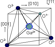
Fig.
12. a) Crystallographic positions of Sr3+, La+, Ga3+
and O2- ions in SLGO lattice; b) Complexes of (Ga-O)1-
arising in SLGO structure (T1 tetrahedron) after g-
or proton irradiation.
The
obtained results can be explained by means of the following process: Ga3+
ion captures the electron which was knocked out from O2- ion by g
or proton irradiation and in a consequence, Ga2+ paramagnetic center
is formed with a spin value equal to S = 1/2.
The process can be illustrated by the following reactions:
O2-+g®O1-
+ e- ; Ga3++e- ®
Ga2+. The measured angular dependences of ESR lines show that this
process inside T1 tetrahedron takes place and suggest that T1 configuration is
energetically the most favorable for this kind of process. Favorable position of
T1 tetrahedra with respect to irradiation process is connected with local
symmetry of the crystal field.
In this
situation the spin Hamiltonian can be written in the following form:
H = g×b×H×S
(2)
where:
S = 1/2, Hrez = n/(g×b/h), g2 = g^2sin2q+g||2cos2q,
b
- Bohr magneton, q
- angle between magnetic field and axis of a centers, h - Planck constant, g -
Lande factor and Hrez - magnetic field.
Because
the direction of an axis each of 4-th (Ga-O)1- complexes inside T1
tetrahedron is of [111] type and maximum Hres is directed along
[110], that is j
= 45o in (001) plane, we have obtained:
g||2 = 2g2[110] -
g2[100] and
g^2
= 2g2[100] - g2[110]
(3)
For
angular dependence ( Fig.7 ) g[100] =2.025
and g[110] = 2.0045 or 2.044 are obtained, and for individual
complex we have obtained g||=1.9838(5) and g^=2.0453(5).
As can
be seen in Fig. 8. the time constant of relaxation process for G1 paramagnetic
defect (part of month - 264 hours) is of the order of a time constant of
relaxation process for the defect
seen in Fig. 1. So, these defects are of the same type and nature.
After g
or proton irradiation
of the SLGO crystal doped with Pr, Dy and Nd, as well as undoped ones, AA bands
appear in the absorption spectra, with maxima at about 270 nm and 370 nm.
The
first band shifts the absorption edge of the crystal towards the longer
wavelengths. This shifting depends on the radiation dose and has similar
character for gammas and protons for doses up to 106 Gy and fluences
up to 1014 protons/cm2. It also depends on the crystal
thickness, which indicates the scattering nature of the produced color centers.
Moreover,
intensity and location of the first maximum, depend on the gamma dose and on the
kind and concentration of a dopant. With the growth of the Pr or Dy
concentration, a maximum of this band decreases, but with the increase of the Nd
concentration, it also increases. The growth of the Pr concentration shift this
maximum towards the shorter wavelengths.
The
second band appears mainly after previous annealing the SLGO crystal in air and
is probably connected with recharging effect of oxygen vacancies that arises in
the crystal during growth process. It leads to decrease of the laser slope
efficiency after thermal annealing process of the laser rod.
Annealing
of the gammas and protons
irradiated crystal with doses up to106 Gy and 1013-1014
particles/cm2, respectively, at 400 oC for three hours,
causes disappearance of the produced color centers.
The
shift of the absorption edge of the irradiated Nd: SLGO crystal can have a
positive influence on its laser properties [7]. Improvement of the laser
emission in a ‘bad’ Nd: SLGO rod at 1,06 mm is probably due to characteristic radiation defect, that is connected
with a greater than in Nd: YAG increase of AA value in the range of absorption
pump spectrum.
Investigations
of ESR spectra show that the centers shifting the absorption edge are of the
paramagnetic origin. Probably, as result of ionizing radiation, paramagnetic
centers are formed according to the reaction Ga3+ + e-®Ga2+.
Received
July 4, 1997; revised October 25, 1997.
[1.] A.
A. Kaminskii, E. L. Belokoneva, B. V. Mill, S. E. Sarkisov and K. Kurbanov,
Phys. Stat. Sol. (a), 97 (1986)
279
[2.] L.
R. Black, D. M. Andrauskas, G. F. de la Fuente, and H. R. Verdun, Proc.
SPIE, 1104 (1989) 175
[3.] W.
Ryba-Romanowski, S. Gołąb, G. Dominiak-Dzik, M. Berkowski, Materials Science and Engineering B, 15(3) (1992) 217
[4.] S. Kaczmarek, Z. Mierczyk, K. Kopczyński, Opto-electronics
Review, 2 (1993) 54
[5.] I. Pracka, W. Giersz, M. Świrkowicz, A. Pajączkowska,
S. Kaczmarek, Z. Mierczyk, K. Kopczyński, Materials
Science and Engineering B, 26(2-3)
1994, 201
[6.] I. Pracka, M. Malinowski, K. Kopczyński, S.
Kaczmarek, Z. Mierczyk, M. Świrkowicz, J.
Kisielewski, T. Łukasiewicz, Proc. SPIE, 3178,
42-44
[7.] S. Kaczmarek, K. Kopczyński, A. Pajączkowska, I.
Pracka, A. O. Matkovskii, Proc. SPIE, 3179,
268-273
[8.] R. Jabłoński, S. M. Kaczmarek, M. Berkowski,
‘Radiation defects in BaLaGa3O7 crystals’, Spectrochimica
Acta Part:A, in print.
S.
KACZMAREK
R. JABŁOŃSKI
I. PRACKA
G.BOULON
T.
ŁUKASIEWICZ
Z. MOROZ
S. WARCHOŁ
K. STĘPKA
Wpływ
promieniowania jonizującego na monokryształy SrLaGa3O7
domieszkowane jonami ziem rzadkich
Streszczenie.
Przedstawiono wyniki badań wpływu kwantów gamma ze źródła 60Co
(1,25 MeV) oraz protonów z akceleratora C30 o energii 26 MeV na absorpcję i
luminescencję monokryształów SrLaGa3O7 domieszkowanych
jonami Nd3+, Dy3+ oraz Pr3+. Centra barwne
powstające po naświetlaniu tych kryształów (dawką 105 Gy oraz 1014
protonów/cm2, odpowiednio), przesuwają krawędź absorpcji w stronę
fal długich o kilkaset nm (dla pręta o długości ok. 36mm). Pokazano również
wyniki badań widm ESR przed i po naświetleniu kwantami gamma. Otrzymano widma
odpowiadające spinowi S=1/2 oraz wartościom: g|| = 1.9838(5) i g^ = 2.0453(5), które mogą być związane z centrami Ga2+
powstałymi w wyniku reakcji przeładowania: Ga3++e-®Ga2+.
Przedstawiono również wyniki badań właściwości generacyjnych kryształów
Nd: SrLaGa3O7, uzyskując poprawę tych właściwości dla
emisji na długości fali 1,06 mm w przypadku silnie zdefektowanego pręta. Wynika to z obecności w tym
krysztale tych samych centrów barwnych, które otrzymano wcześniej w wyniku naświetlania
tego kryształu kwantami gamma lub protonami (centra Ga2+).